
Synopsis
SARS CoV-2 is affecting millions of people all over the world. Clinical studies find out SARS CoV-2 neurological involvement. Although respiratory symptoms are more common, the virus affects also the Central and Peripherical Nervous System. Preliminary studies demonstrates that Structural Magnetic Resonance Imaging and Electroencephalograms can be precious allies for the detection of SARS CoV-2. To better understand the middle and long term impact of SARS CoV-2 on neurological activity, Functional Magnetic Resonance Protocol should be included.
Abbreviations: New coronavirus 2019: SARS CoV-2, Infection caused by new coronavirus 2019: COVID-19, Electroencephalography: EEG, Magnetic Resonance Image: MRI.
COVID-19 effects on the Nervous System
Clinical evidence of neuro-infection caused by coronaviruses has been already discovered by clinical studies dealing with Severe Acute Respiratory Syndrome (SARS CoV) and Middle East Respiratory Syndrome (MERS CoV)1–3. After the spreading of the new coronavirus SARS CoV-2 all over the world, its neurological effect has been supposed to be similar to the other coronaviruses and preliminary studies performed since January 2020 seem to confirm this hypothesis4,5. Mao and colleagues carried out astudy on a group of 214 patients with a serious form of the infections enrolled in Wuhan, they have demonstrated that SARS-CoV2 acts on both Central Nervous System (CNS) and Peripherical Nervous System (PNS)6.
In CNS, COVID-19 is associated to large amount of diseases as encephalopathy, meningitis, ischemic and hemorrhagic stroke7. Preliminary studies confirmed the interactions between the virus and CNS, with the most severe complications in patients with already assessed neurological disease. They have been demonstrated that the SARS-CoV2 influences the cytokine production, causing the development of encephalopathies and the possible breakdown of blood-brain-barrier8,9. COVID-19 effect on the PNS led to dysfunction of smell and taste (i.e. anosmia, ageusia), muscle injury and post-infective neuropathies as the Guillain-Barre syndrome7. Although the most critical neuro-pathologies have been recorded in patients with already pre-existent disease10, it is fundamental to investigate the COVID-19 neuro-effects in order to be able to detect the virus infection also through the analysis neurological symptoms.
How to detect COVID-19 neurological effects
The experience of previous investigations on the other coronaviruses tells us that in order to investigate whether COVID-19 infections modify the activity of cerebral regions, functional MRI (fMRI) and Electroencephalography (EEG) can be very useful to assess the mid/long-term effects of the virus on the CNS. High resolution Magnetic Resonance Images (MRI) are considered the gold standard to investigate the lesions of cerebral tissues and possible cerebral hemorrhages11, therefore, different MRI techniques have been already employed in preliminary studies about the relationship between COVID-19 and CNS injury12.
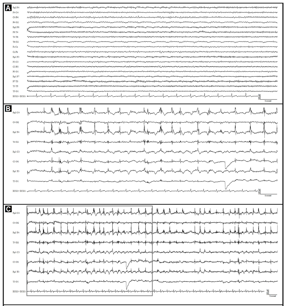
In patients with previously diagnosed CNS pathologies and positive to SARS-CoV2, the virus seems to increase neurological disorder manifestation, even without causing new brain damage or bleeding events. Vollono et al, 2020 reported the clinical case of a woman with postencephalitic epilepsy presenting a focal status epilepticus after the primary presentation of COVID-1913. EEG performed during a follow-up resulted normal, while the EEG performed during the recovery period after COVID-19, revealed irregular delta activity predominantly in fronto-centro-temporal regions (Figure 1). In this case brain MRI has not shown new injuries.
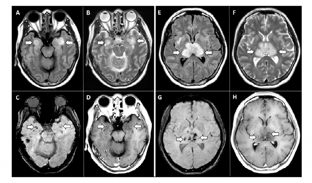
Poyiadji et al, 2020 reported the case of a woman on late fifties with severe acute respiratory syndrome associated to COVID-19 and an altered mental status14. Structural MRI has been used to detect the presence hemorrhagic events. Figure 2 shows the coronal section of the MRI. T2 FLAIR shows the hyperintense regions in correspondence of medial temporal lobes (refer to panel A and B) and of thalami (panel E and F). The hemorrhagic event results in hypointense region in susceptibility weighted images (panel C and G) with the rim enhanced in the post-contrast images (panel D and H). Lesions in the thalamic and medial lobe area are a typical feature of Acute Necrotizing Encephalopathy (ANE). Other characteristic brain structures that might be affected by ANE are brainstem and cerebellum15. Therefore, in a future study, it would be useful to carry out the analysis of these areas to evaluate the presence of damages in COVID-19 patients and to investigate long terms effects of the virus on brain activity16.
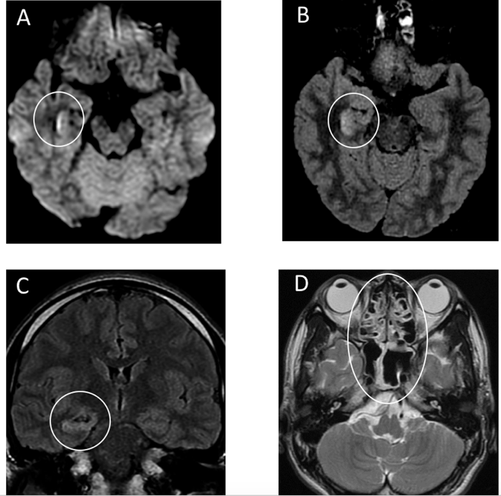
Patients affected by SARS-CoV2 might result negative to the nasal swab but present anyway the neurological diseases induced by the virus as headache, confusion and, in the most severe case, loss of consciousness. Moriguchi et al, 2020 reported the case of a 24 years male with fever, cough and headache whose COVID-19 nasal swab resulted negative17. He has been recovered in Intensive Care Unit (ICU) few days after the swab because he presented sever consciousness disturbance and seizure episode. Cerebro-Spinal Fluid (CSF) has been tested showing the presence of COVID-19. Brain MRI has been performed to detect the presence of cerebral lesions. Imaging features showed in Figure 3) lead to the diagnosis of right lateral ventriculitis and encephalitis located in right mesial lobe and hippocampus.
EEG and MRI abnormal findings of the studies mentioned above demonstrate that encephalopathies and brain lesion might be considered as symptoms of COVID-19 at the same level of acute respiratory syndrome. In addition, these findings suggest that neurological symptoms are not characteristic of a specific age range but, in presence of a severe form of COVID-19, they can manifest both in young and old patients13,14. In the past, other coronaviruses have been found responsible of neurological disease and assessed the importance of brain imaging technique to investigate the brain areas targeted by the infection18. Therefore, MRI technique should be considered in the protocol of diagnosis for suspected COVID-19 patients with neurological symptoms.
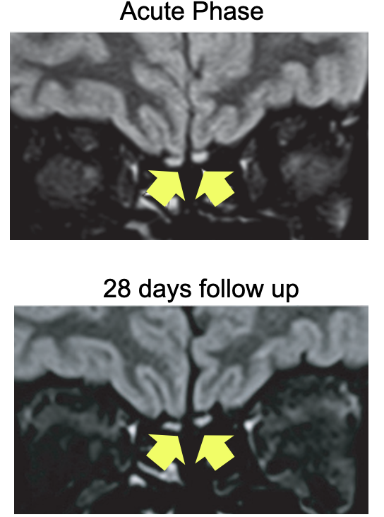
From a cellular-scale point of view, the COVID-19 gateway seems to be the olfactory epithelium, where the cellular factors for the virus entry lie. Their absence in the olfactory sensory neurons makes the COVID-19 induced loss of olfaction (anosmia) not fully understood19. Past studies proposed MRI technique to investigate whether anosmia causes morphological changes of olfactory bulbs20,21. Politi et al, 2020 repots the clinical case of a 25-year-old female positive to COVID-19 with persistent sever anosmia. The brain MRI shows an hyperintense signal in correspondence of olfactory bulbs (Figure 4). A follow-up MRI performed about one month later revealed that the olfactory bulbs were thinner and slightly less hyperintense22. This finding suggests that performing MRI longitudinal studies focused on olfactory bulbs may be useful to assess for possible temporal evolution from the acute to the follow-up phase23. It could be very interesting to investigate the effects of COVID-19 on the olfactory system using high resolution MRI technique focused on the olfactory bulbs. Furthermore, performing high contrast and high spatial resolution MRI can be a powerful tool to investigate COVID-19 effect not only on CNS but also on PNS.
In conclusion, considering the similarity between SARS-CoV and SARS CoV-2, it is needed to be clarified how this last one affects Nervous System, as well as the respiratory tract, and if this infection may be partially responsible for the acute respiratory failure of patients with COVID-19.
In addition, we remark the importance of structural MRI techniques to detect possible lesions or hemorrhages. An ad hoc protocol of high-resolution MRI of these structures could be defined to collect data on the COVID-19 and to compare the findings with previous coronaviruses (SARS CoV, MERS) effects on CNS. Furthermore, since the evidences mentioned above underline the importance of MRI in investigation of COVID-19 effects on CNS, we are wondering on the possibility to insert Functional MRI (fMRI) in research protocol on COVID-19. The studies performed until now have not reported fMRI or EEG acquisitions as a diagnostic tool. The reason could be found in the need to act very quickly during the peak phase of the health emergency caused by COVID-19.
These clinical exams allow to investigate brain activity in spatial and temporal domain respectively. In addition, Blood Oxygenation Level Dependent signal (BOLD) is strictly related to neurovascular system10, therefore it could be investigated whether the virus effect on neurovascular system is reflected in BOLD signal.
The definition of a follow-up protocol for patients affected by COVID-19 with neurological symptoms, including both structural and functional MRI and EEG, would allow to perform longitudinal study on cerebral lesions and the analysis of the changes occurred on the cerebral activity of patients recovered from COVID-19, who showed neurological symptoms associated to the virus. Furthermore, as mentioned in Vollono et al, 2020 study, in the case of COVID-19 patients already under follow-up for previously assessed CNS diseases, the acquisitions of fMRI and EEG allow the investigation of COVID-19 effect on an already compromise CNS.
References:
1. Lau K-K, Yu W-C, Chu C-M, et al. Possible central nervous system infection by SARS coronavirus [Internet]. Emerg. Infect. Dis. 2004;10(2):342–344.Available from: https://pubmed.ncbi.nlm.nih.gov/15030709
2. Bergmann CC, Lane TE, Stohlman SA. Coronavirus infection of the central nervous system: host-virus stand-off. Nat. Rev. Microbiol. 2006;4(2):121–132.
3. Weiss SR, Leibowitz JL. Coronavirus pathogenesis. Adv. Virus Res. 2011;81:85–164.
4. Lu L, Xiong W, Liu D, et al. New‐onset acute symptomatic seizure and risk factors in Corona Virus Disease 2019: A Retrospective Multicenter Study. Epilepsia 2020;0–1.
5. Carod-Artal FJ. Neurological complications of coronavirus and COVID-19. Rev. Neurol. 2020;70(9):311–322.
6. Mao L, Jin H, Wang M, et al. Neurologic Manifestations of Hospitalized Patients with Coronavirus Disease 2019 in Wuhan, China. JAMA Neurol. 2020;77(6):683–690.
7. Koralnik IJ, Tyler KL. COVID-19: A Global Threat to the Nervous System. Ann. Neurol. 2020;88(1):1–11.
8. Wang D, Hu B, Hu C, et al. Clinical Characteristics of 138 Hospitalized Patients with 2019 Novel Coronavirus-Infected Pneumonia in Wuhan, China. JAMA – J. Am. Med. Assoc. 2020;323(11):1061–1069.
9. Health LLIJ of M and, 2020. COVID-19: A Systematic Review of The Mechanisms of Injury to The Central Nervous System This Far [Internet]. Iajmh.Com [date unknown];Available from: https://www.iajmh.com/iajmh/article/view/115%0Apapers3://publication/uuid/3EF28B73-E4DA-4405-B2EF-FCAB6303C64E
10. Bridwell R, Long B, Gottlieb M. Neurologic complications of COVID-19 [Internet]. Am. J. Emerg. Med. 2020;38(7):1549.e3-1549.e7.Available from: https://doi.org/10.1016/j.ajem.2020.05.024
11. Thiebaut de Schotten M, Croxson PL, Mars RB. Large-scale comparative neuroimaging: Where are we and what do we need? [Internet]. Cortex 2019;118:188–202.Available from: https://doi.org/10.1016/j.cortex.2018.11.028
12. Asadi-Pooya AA, Simani L. Central nervous system manifestations of COVID-19: A systematic review [Internet]. J. Neurol. Sci. 2020;413:116832.Available from: https://pubmed.ncbi.nlm.nih.gov/32299017
13. Vollono C, Rollo E, Romozzi M, et al. Focal status epilepticus as unique clinical feature of COVID-19: A case report [Internet]. Seizure 2020;78(April):109–112.Available from: https://doi.org/10.1016/j.seizure.2020.04.009
14. Poyiadji N, Shahin G, Noujaim D, et al. COVID-19–associated Acute Hemorrhagic Necrotizing Encephalopathy: CT and MRI Features [Internet]. Radiology 2020;201187.Available from: https://doi.org/10.1148/radiol.2020201187
15. Narra R, Mandapalli A, Kamaraju SK. Acute necrotizing encephalopathy in an adult. J. Clin. Imaging Sci. 2015;5:20.
16. D’Angelo E. Physiology of the cerebellum [Internet]. 1st ed. Elsevier B.V.; 2018.Available from: http://dx.doi.org/10.1016/B978-0-444-63956-1.00006-0
17. Moriguchi T, Harii N, Goto J, et al. A first case of meningitis/encephalitis associated with SARS-Coronavirus-2 [Internet]. Int. J. Infect. Dis. 2020;94:55–58.Available from: https://doi.org/10.1016/j.ijid.2020.03.062
18. Li YC, Bai WZ, Hashikawa T. The neuroinvasive potential of SARS-CoV2 may play a role in the respiratory failure of COVID-19 patients. J. Med. Virol. 2020;92(6):552–555.
19. Baig AM, Khaleeq A, Ali U, Syeda H. Evidence of the COVID-19 Virus Targeting the CNS: Tissue Distribution, Host-Virus Interaction, and Proposed Neurotropic Mechanisms. ACS Chem. Neurosci. 2020;0–3.
20. Chung MS, Choi WR, Jeong HY, et al. MR imaging-based evaluations of olfactory bulb atrophy in patients with olfactory dysfunction. Am. J. Neuroradiol. 2018;39(3):532–537.
21. Prasanth P, Elton M L. Clinical Image: Olfactory Bulb and Tract Aplasia/Hypoplasia on MRI. Otolaryngol. Res. Rev. 2017;1(1):34–35.
22. Politi LS, Salsano E, Grimaldi M. Magnetic Resonance Imaging Alteration ofthe Brain in a Patient With Coronavirus Disease 2019 (COVID-19) and Anosmia. JAMA Neurol. 2020;
23. Mahboobeh Karimi Galougahi, Jahangir Ghor- bani, Mehrdad Bakhshayeshkaram, Ali Safavi Naeini SH. Letters to the Editor Olfactory Bulb Magnetic. 2020;(January):1–2.
You may also like this

Europe’s top universities: Where research thrives in 2025

Microbes and Mood: Deciphering the Connection for a Happier You!
Can’t solve a maze? Ask a slime mould!

The neurobiology behind the Christmas spirit
