
by Giuseppe Scarlata, Domenico Gangemi, Maria Chiara Rosace, Antonino Napoleone | November 07, 2020
The scientific journal IDCases is well known for the publication of case reports dedicated to infectious diseases, and last June, one in particular entitled “COVID-19 and Plasmodium Vivax malaria co-infection” aroused considerable clinical interest. The authors, belonging to the Department of Internal Medicine of the Hamad Medical Corporation (Doha, Qatar) presented a unique case report of a patient suffering from malaria and COVID-19 co-infection, to whom antimalarial drugs were administered. This is the first case reported in the literature. The infection caused by SARS-CoV-2 needs no further presentation as it has been extensively investigated in previous articles (read more: Covid-19 – Asymptomatic cases and incubation period, Covid-19 – Was the virus truly created in the laboratory? and Asymptomatic cases and incubation period), on the contrary regarding malaria, it is necessary to explain a bit about the pathogen and its biological cycle.
What is malaria?
Malaria is a systemic infection caused by parasites belonging to the genus Plasmodium. The hematophagous female mosquito belonging to the genus Anopheles is the definitive host of these parasites. The mosquitos are capable of infecting humans, who are the intermediate host of the parasites. The parasite biological life cycle occurs therefore between the human and the mosquito hosts. Its life cycle is divided into three clearly distinguished phases: the first two occur within the human hosts (hexo- eritrocitary and erythrocytic schizogony cycles) while the last one within the mosquito (sporogonic cycle). The mosquito will then inject the plasmodium within the intermediate host, favouring the beginning of a new biological cycle (Figure 1)
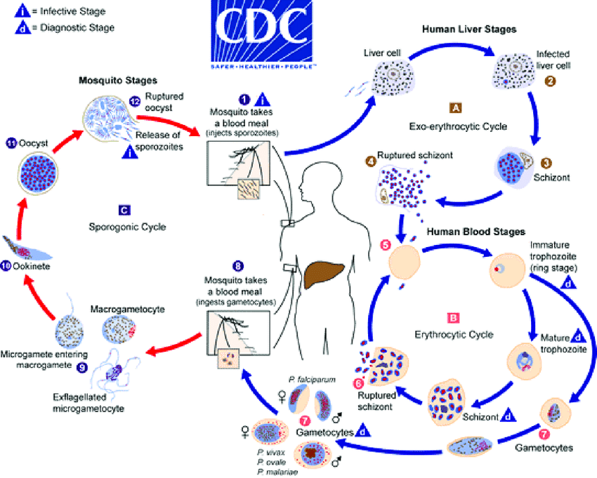
Figure 1. During the blood meal (1) a mosquito infected female inoculates sporozoites in the human host [A]:
Sporozoites reach the liver and infect liver cells (2) where they multiply and mature into schizonts within 7-21 days (incubation period). Schizonts rupture and release thousands of merozoites which invade the bloodstream. In P. vivax and P. ovale, some sporozoites do not immediately mature into schizonts but remain in a dormant stage (hypnozoites) before continuing the cycle and maturing into schizonts. These are the cause of relapses weeks, or even years later.
Erythrocytic schizogony cycle (in humans) [B]: Merozoites infect red blood cells (5). The ring stage trophozoites mature into schizonts, which rupture releasing merozoites (6) which invades new red blood cells. The erythrocytic cycle lasts for 24 hours in P. knowlesi , 48 hours in P. falciparum, P. vivax, P. ovale and 72 hours in P. malariae. Following many cycles, some parasites differentiate into sexual erythrocytic stages (gametocytes) (7). Blood stage parasites are responsible for the clinical manifestations of the disease. The gametocytes, male (microgametocytes) and female (macrogametocytes), are ingested by the mosquito during a blood meal (8)
Sporogonic cycle (mosquito) [C]: in the mosquito’s stomach gametocytes mature to gametes. Microgametes exflagellate and fertilise macrogametes forming the zygote (9). The zygotes in turn become motile and elongated (ookinetes) (10). They penetrate the stomach wall and develop into oocysts (11). The oocysts grow, rupture, and release sporozoites that reach the mosquito’s salivary glands (12). The inoculation of sporozoitis into a new human (1) perpetuates the malaria life cycle. The duration of the sporogonial cycle varies depending on the Anopheles species, temperature and humidity, but in general, it lasts for 8-16 days.
The species capable of infecting humans are Plasmodium malariae, P. falciparum, P. knowlesi, P. ovale and P. vivax. Each of them, depending on the duration of the erythrocyte cycle (24-72 hours) may cause benign or malignant febrile episodes every other day (except for P. knowlesi which causes daily fever). Currently, malaria is endemic in 87 countries worldwide. Among these, the African continent is certainly the most plagued one, particularly the area of central and sub-Saharan Africa, where P. falciparum is predominant. The plan of the World Health Organization (WHO) is to eradicate the disease in 35 other countries by 2030 by investing funds already this year to implement research and development of therapeutic strategies, prevention and vaccination and surveillance programs. However, it is needless to say that the current historical situation worldwive leaves no margin for such good intentions.
The clinical case reported in the literature
Here is reported the case of a 34-year-old man with no previously diagnosed pathologies presented to the hospital with a three-day history of fever (39.6°C), with myalgia and vomiting. No respiratory symptoms were shown. The man mentioned a trip to Pakistan three months prior to develop the current symptomatology.
Therefore, the clinical picture at the hospitalization was:
- Febrile state (39.6°C) with vomiting and abdominal pain
- Blood pressure 109/72 mmHg
- Tachycardia (148 beats per minute)
- Tachypnea (respiratory rate of 24 respiratory breaths per minute)
- Oxygen saturation of 97 %
Physical examination revealed a jaundiced man (yellowish skin discolouration caused by the increase of bilirubin in the blood), although ultrasound of the abdomen revealed hypoechoic liver with prominent periportal tracts and no evidence of cholelithiasis, i.e. gallstones in the gallbladder lumen. A further radiological examination by chest X-ray showed negative results, while the electrocardiogram showed sinus tachycardia. Subsequently, haemato-chemical tests were performed and reported:
- Leukopenia: white blood cell count 3.6 × 10³/μL (reference range 4.0-10.0 × 10³/μL)
- Lymphopenia: 0.2 × 10³/μL (reference range 1.0-3.0 × 10³/μL)
- Haemoglobin 14.5 g/dL (reference range 13.0-17.0 g/dL)
- Thrombocytopenia with platelet count 30 × 103/mL (reference range 150-400/mL)
- Direct hyperbilirubemia
- C reactive protein 238 mg/l (reference range 0-5 mg/l)
- Procalcitonin 61.30 ng/L (reference range 0,5-2,0 ng/L)
- Lactic acid 2.4 mmol/L (reference range 0.5-2.2 mmol/L)
- Ferritin 1.831,0 μg/L (reference range 48-420 μg/L)
- D-dimer 5,40 mg/L (reference range 0,00-0,44 mg/L)
Following the performed investigations, the etiological suspicion was oriented to claim it as a SARS-CoV-2 infection (supported by febrile state, haematochemical tests showing leukopenia, lymphopenia, and increased values of PCR, PCT and D-dimer) on one hand and by a parasitic infection, malaria in this case (supported by thrombocytopenia, direct hyperbilirubinemia, jaundice, fever for three days, trip to Pakistan) on the other hand. Subsequent laboratory tests were then performed, including the malaria smear on blood sample (Figure 2) and the Real-Time PCR (Polymerase Chain Reaction) on nasopharyngeal swab.

The first one is a highly specific test for the diagnosis of species of the genus Plasmodium. It allows the calculation of the parasitemia (% of red blood cells infected by the parasite). In this case, the patient tested positive for Plasmodium vivax with a significant parasitemia of 1.2%. RT-PCR on nasopharyngeal swab revealed a concomitant positive result for SARS-CoV-2. The initial therapy involving Arthemeter/lumefantrine (Coartem®) was initiated with fluid resuscitation, but it was not tolerated by the patient due to vomiting. It was therefore immediately replaced with intravenous artesunate at a dose of 2.4mg/kg at 0, 12, 24, 48 hours; once the patient was able to tolerate oral therapy, anti-malarial treatment was continued with artemether-lumefantrine. The mechanism of action of these drugs involves a biochemical process that leads to the production of oxygen free radicals which damage the parasite and cause its death.
It should be noted that for the entire duration of the hospital stay the patient did not show any respiratory symptoms so oxygen supplementation was not required. New tests performed after the treatment reported normalization of the liver parenchyma, normal white blood cell count (6.8 × 10³/μL), improved platelet count (144 × 103/μL) and total bilirubin 1.35 mg/dL. Repeated malaria smear after 5 days from initial presentation revealed clearance of parasitemia, while RT-PCR on nasopharyngeal swab repeated seven days later was confirmed as negative. Upon discharge, the patient was placed in a facility to complete a 14-day isolation and, according to protocol, and a primaquine prescription was given orally for the duration of the isolation to treat any relapse.
In conclusion
The authors have more doubts than certainties, but promptly formulated various hypotheses:
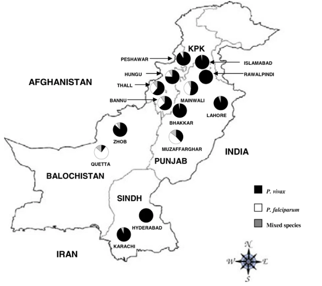
- The source of infection is suspect: is it a local transmission, or is it a reactivation of parasites at the hypnozoite stage (see Figure 1) localized in the liver, following an infection contracted in the previous months? The latter hypothesis seems to be the most likely, considering the fact that Qatar is not an endemic area for malaria, like Pakistan (Figure 3), where the patient travelled three months earlier.
- It is essential that the scientific community adopts an accepted and validated therapeutic program to treat COVID-19, as currently, many drugs are still in more or less promising clinical trials (read more: Which drugs are available against SARS-CoV-2).
- Has anti-malarial therapy effectively eradicated SARS-CoV-2 infection as well? Currently, the hypothesis seems to be likely, due to the fact that the anti-malarial Chloroquine/Hydroxychloroquine has already been used as a therapeutic approach in patients with COVID-19, in order to inhibit virus-target cell fusion.9.
The therapeutic approach based on Chloroquine / Hydroxychloroquine for the treatment of COVID-19 is currently being studied in more than 80 registered clinical trials. Several in vitro studies have shown that these drugs have a consistent anti-viral effect against Coronavirus. However, to date, no studies have been published in which the use of Chloroquine/Hydroxychloroquine would be beneficial for the prophylaxis and/or treatment of COVID-19 patients. Moreover, the administration of these drugs is associated with cardiotoxicity, myopathies and retinopathies. A recently published study in Nature even denied the validity of this approach, since the use of Chloroquine / Hydroxychloroquine was not effective in blocking SARS-CoV-2 infection in human lung cells, excluding any protective and preventive effect against COVID-19. However, new clinical trials involving new drugs are needed more than ever, it is never known that these hypotheses can be confirmed, but in the meantime, it is good not to sponsor therapies not validated yet or create false illusions among the population. Share and have faith in medical science rather than the media!
References:
- Sardar S, Sharma R, Alyamani TYM, Aboukamar M. COVID-19 and Plasmodium vivax malaria co-infection. IDCases. 2020, 21, e00879. doi: 10.1016/j.idcr.2020.e00879
- CDC (Centre for Diseases Control and Prevention). Malaria. CDC. https://www.cdc.gov/parasites/malaria/index.html Updated August 20, 2020. Accessed November 4, 2020.
- World Malaria Report, 2019 [WHO].
- Khattak AA, Venkatesan M, Nadeem MF et al. Prevalence and distribution of human Plasmodium infection in Pakistan. Malar J. 2013, 12,297. doi: 10.1186/1475-2875-12-297.
- Hoffmann, M., Mösbauer, K., Hofmann-Winkler, H. et al. Chloroquine does not inhibit infection of human lung cells with SARS-CoV-2. Nature 585, 588–590 (2020). https://doi.org/10.1038/s41586-020-2575-3.
- Khuroo MS. Chloroquine and hydroxychloroquine in coronavirus disease 2019 (COVID-19). Facts, fiction and the hype: a critical appraisal. Int J Antimicrob Agents. 2020;56(3):106101. doi:10.1016/j.ijantimicag.2020.106101.
You may also like this

COVID-19 – What is the CT value and what does it convey about infectivity?

COVID-19 and vaccines: Can the transmission of the virus be stopped?

Covid-19 – Vaccines options available: how to choose?

Covid-19 – Recovery in symptomatic and asymptomatic patients: post-COVID-19 Syndrome
